Learning Outcomes
- State the components of the cell theory.
- State the importance of multicellularity.
- Distinguish the domains under Prokarya and differentiate them from Eukarya
- Identify the components and describe the function of cellular components in plant cells
- Identify the components and describe the function of cellular components in animal cells
- Identify the components and describe the function of cellular components of Amoeba
- Cell Membrane, Contractile vacuole, cytoplasm, food vacuole, nucleus, nucleolus, pseudopod, ribosome
- Identify the components and describe the function of cellular components of Euglena
- Cell Membrane, Chloroplast, Contractile vacuole, cytoplasm, flagellum, nucleus, nucleolus, pseudopod, ribosome, stigma
- Name the three main layers of the plant cell wall, and list the chemical components of each layer.
- Describe the general structure and function of the nucleus.
- Describe the basic structure and function of each of the following cell components: cytoplasm, vacuole, ribosome, endoplasmic reticulum, Golgi complex, lysozome, peroxisome, chloroplast and mitochondrion.
- Describe the origins of Chloroplasts and Mitochondria
- List the components of the cytoskeleton, and describe its general role in the cell.
Presentation
The Study of Cells https://h5p.org/node/273473
Worksheet
https://github.com/jeremyseto/bio-oer/blob/master/figures/cellstructure/Cell-Worksheet.pdf
Parts of Cells

Ring of life diagram illustrating the divergence of prokaryotes and the subsequent endosymbiosis that gave rise to eukaryotes. Credit: Maulucioni [CC-BY-SA 3.0]
All living things on the planet share common features in their parts. The ring of life illustrates the diversification of life from prokaryotic domains and the reincorporation into more complicated cells of the domain eukarya.
Cell membrane

The cell membrane is the barrier that separates the cytoplasm from the external world. The cell membrane consists primarily of phospholipids in a bilayer. Phospholipids are amphipathic with a polar head (phosphate group) and a hydrophobic tail (2 hydrocarbon chains). Due to the chemical properties of the heads being attracted to water and the tails having a desire to avoid water, phospholipids self assemble into micelles. Cell membranes form from a phospholipid bilayer where the lipid tails interact with each other and the phosphate heads face the external water environment or the internal cytoplasm of the cell.
The cell membrane does not solely consist of phospholipids but also have proteins and cholesterol inserted into the bilayer. As the image of the bilayer above indicates, the molecules are constantly moving and flow in a lateral motion. Cholesterol modulates the fluidity of this motion. Proteins associated with the membrane may sit on either side (peripheral proteins) of the membrane or pass through both layers of the membrane (transmembrane proteins). The model that describes the components of the cellular membrane is referred to as the Fluid Mosaic Model. This model states that the cell membrane is a mosaic of 1)Phospholipids 2)Proteins 3) cholesterol that move about in a side to side motion.

Cytoplasm

Cytoskeleton

The cytoplasm consists of all components of the the cells enclosed by the cell membrane. The cytosol is a component of the cytoplasm that corresponds to the intracellular fluid matrix. This cytoplasmic environment is a dense matrix of proteins and other molecules.
Ribosomes

The ribosome is an enzymatic complex composed of protein and RNA (ribosomal RNA or rRNA). Ribosomes consist of two subunits, the large and the small subunits. The large and small ribosomal subunits assemble together to translate genetic information from messenger RNA (mRNA) into proteins. The subunits are so large that they are described by their sedimentation in a centrifuge in S units (Svedberg unit). Complete prokaryotic ribosomes are 70S and consist of a 50S large subunit and 30S small subunit. While they function in a similar manner, eukaryotic ribosomes are larger at 80S from the 60S large subunit and 40S small subunit. The differences in ribosomes between prokaryotes and eukaryotes make them a suitable target for antibiotics like streptomycin.

An interlinking network of proteins extends throughout the cytoplasm of cells to provide structure and mobility. These proteins polymerize from small subunits and are categorized into 3 major categories based on their size: microfilaments, intermediate filaments and microtubules.
- http://pdb101.rcsb.org/motm/175
- https://pdb101.rcsb.org/motm/19
- https://youtu.be/YTv9ItGd050
- https://youtu.be/tO-W8mvBa78
Undulipodia

Cells can propel themselves through their environment using undulipodia (unduli – swing, podium – foot). These structures are extracellular extensions of cytoskeleton called cilia or flagella. Flagella are often long and exist alone or in pairs while cilia are short and numerous. They have a distinctive organization 9+2 where a central doublet of microtubules is surrounded by 9 doublets of microtubules. Motor proteins are associated with this microtubule organization to provide the movement. Analogous structures exist in prokaryotes as well, with a slightly different organization and means of propulsion.
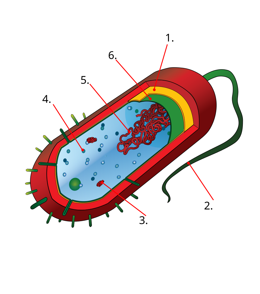
Prokaryotic Cell
Diversity of Prokaryotic Cells

Prokarya (pro – before, karya – nucleus) is the superdomain containing Bacteria and Archaea. Prokaryotes do not possess nuclei, however chromosomal DNA is present in a nucleoid region of the cytoplasm. In addition to the genomic DNA, small mobile DNA elements called plasmids may exist in some bacteria that provide additional genetic diversity.
Bacteria have two common shapes: spherical (cocci) or rod-shaped (bacili). Other morphologies like filamentous and spiral (spirochete) exist, as well as more complicated shapes.
Cell Walls of Bacteria
Cell walls of bacteria exist immediately outside of the cell membrane and protect the cell from internal turgor pressure. These cell walls are composed of a carbohydrate called peptidoglycan. The cell walls of bacteria are relatively porous and not a permeability barrier. Because the cell wall provides rigidity and structure, compromising the integrity is detrimental to the cell. Penicillin is an antibiotic called a β-Lactam that interferes the cross-linking of the peptidoglycan and can effectively control bacterial populations.
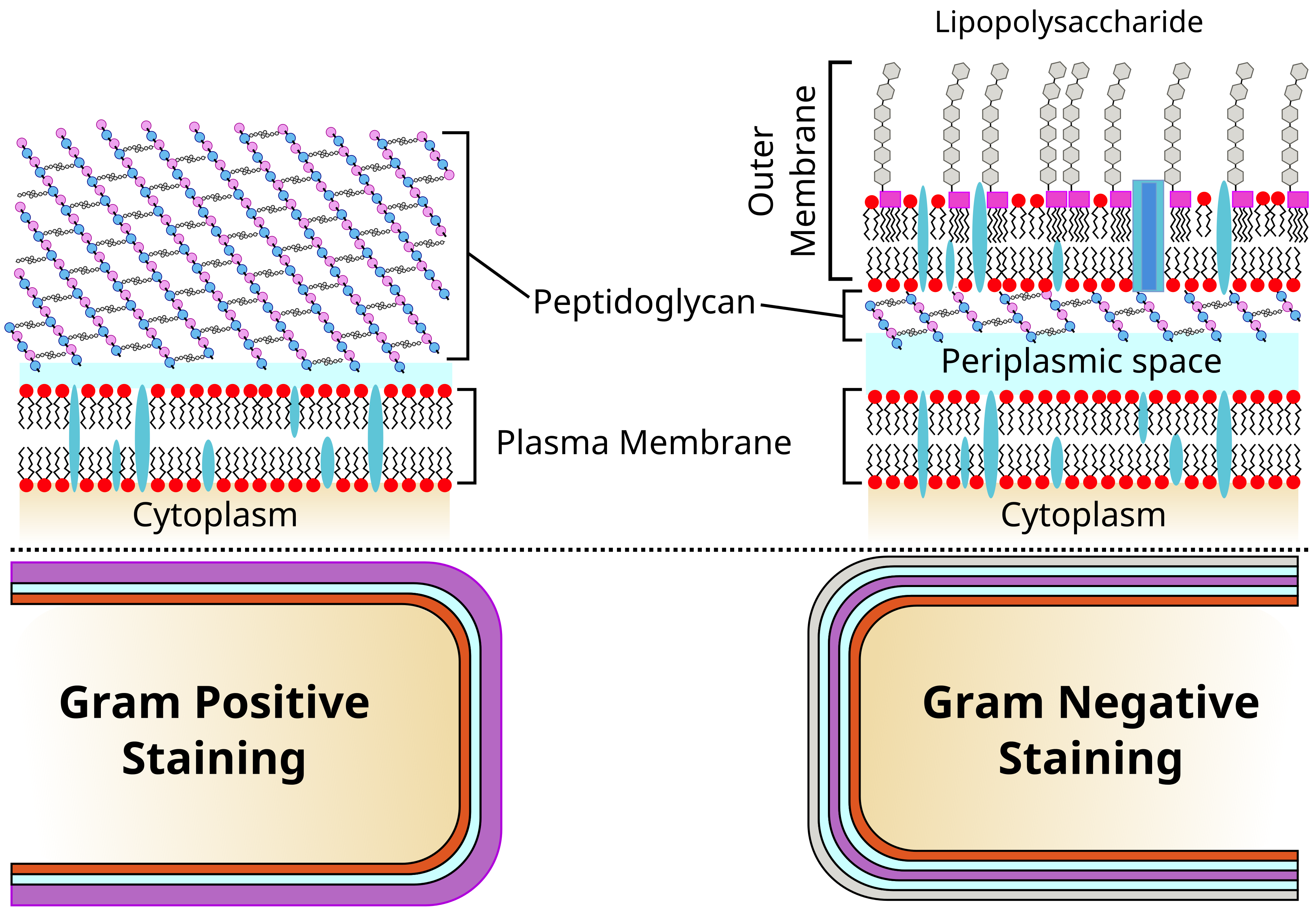
Bacteria are generally separated into two categories based on their cell wall composition. The Gram stain is a methodology that detects the peptidoglycan by a dye called crystal violet. Gram-positive cells stain purple because of the thick layer of peptidoglycan at the surface of the cell. Gram negative cells stain pink with a counterstain since the crystal violet can not access the thin layer of peptidoglycan beneath an outer membrane of lipopolysaccharides. β-Lactams are less effective on Gram-negative cells due to these structural differences. Properties of the outer membrane in Gram-negative bacteria vary greatly between species and render antibodies less effective in some cases.

Eukaryotic Cells
Eukarya (eu – true, karya- nucleus) is the domain of cells defined by the containment of the genomic DNA in a nucleus.
Nucleus and Nucleolus

Eukaryotic nuclei enclose the genomic DNA in a double membrane derived from the endomembrane system called the nuclear envelope. Because the nucleus houses the genetic information set to run the cell, it is often referred to as the “brain” of the cell. Transport of molecules between the cytosol and nuclear matrix occur through nuclear pores within the nuclear envelope. Some cells exhibit a noticeable darker portion of the nucleus called the nucleolus. The nucleolus is an area of the nucleus devoted to ribosome biogenesis.

Endomembrane system

Golgi Apparatus

https://commons.wikimedia.org/wiki/File:YeastGolgiMovieeLifec.ogv Effrosyni Papanikou Kasey J Day Jotham Austin II Benjamin S Glick. Yeast golgi dynamics. Green labels early golgi, red labels late golgi. From https://dx.doi.org/10.7554/eLife.13232 eLife 2015;4:e13232
Cytoplasmic organelles
Endosymbiosis

Labeling Exercises
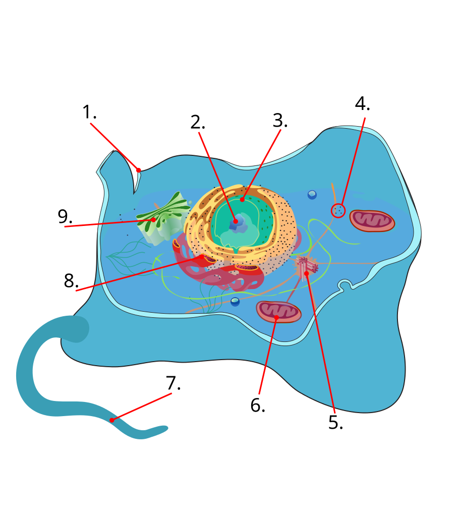
Animal Cell
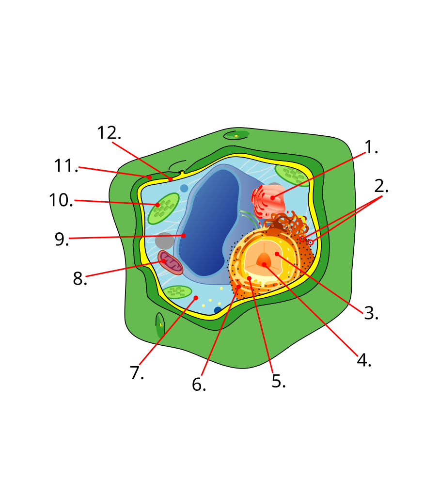
Plant Cell
Cell Walls


This entry is licensed under a Creative Commons Attribution-NonCommercial 4.0 International license.