Contents
Learning Outcomes
- Identify and describe the function of the primary parts of a binocular microscope.
- Calculate total magnification with each objective
- Demonstrate basic skills of light microscopy: locating and bringing into focus, using the correct procedure, an object under low and high power.
The Light Microscope
Hooke’s Cell
In 1665, Robert Hooke published Micrographia, a book that illustrated highly magnified items that included insects and plants. This book spurred on interest in the sciences to examine the microscopic world using lenses but is also notable for Hooke’s observations of cork where he used the word “cell” in a biological sense for the first time.The father of Microbiology: van Leeuwenhoek
The Dutch tradesman Antonie van Leeuwenhoek used high power magnifying lenses to examine the parts of insects and to examine the quality of fabric in his drapery business. He began to experiment with pulling glass to generate lenses and developed a simple microscope to observe samples. Using a simple single lens with a specimen mounted on a point, he was able to identify the first microscopic “animalcules” (little animals) that will be later known as protozoa (original animals).Though van Leeuwenhoek’s apparatus was simple, the magnifying power of his lenses and his curiosity enabled him to perform great scientific observations on the the microscopic world. He was ridiculed for fabricating his observations of protists at first. Ever the scientist, van Leeuwenhoek examined samples of his own diarrhea to discover Giardia intestinalis. While he did not make the connection of the causative nature of this microorganism, he described the details of the way this organism could propel itself through the medium in great detail.Modern Compound Microscope
Unlike van Leeuwenhoek’s single lens microscope, we now combine the magnifying power of multiple lenses in what is called a compound microscope.
Parts of a Compound Light Microscope
- Base: bottom part of the microscope stabilizes it and allows it to stand upright.
- Arm or stand: connects the base to the Binocular Tube.
- Body Tube or Binocular Tube: contains the lens systems and mirrors that allows the specimen to be seen and magnified.
- Ocular Lens or Eyepiece: the lens found at the top of the binocular tube, through which the eye observes the specimen. A monocular microscope would have one ocular lens; a binocular microscope has two ocular lenses. By looking through the ocular lens at times a pointer can be seen. The pointer can be used to point to a specific structure in the specimen. An ocular lens may also have an ocular micrometer, an arbitrary ruler used to measure the length of specific structures in the specimen. Most ocular lenses have a magnification or 10X.
- Objective Lenses: these are the lenses closest to the specimen being studied and found on the revolving nosepiece. Depending on the microscope the objective lenses can have a magnification of 4X, 10X, 40X and 100X, by rotating the nosepiece the observer can choose a different lens and change the magnification used to observe the specimen.
- Mechanical stage – flat surface on which the slide is held in place by the specimen holder. The stage can be moved up or down by the Coarse adjustment knob or Fine adjustment knob which are on the microscope arm to focus on different levels of the specimen observed. The mechanical stage may also be moved back and forth or side to side using the Mechanical Stage Coaxial Control Knobs or x-axis knob and y-axis knob.
- Condenser: The light path is from the in-base illuminator through air to the condenser mount that contains the diaphragm lever (or Iris) and the condenser, through the specimen mounted on a glass slide, through air again and finally through the optical system of objective lens, mirrors, and ocular lenses in the tube. The iris diaphragm regulates the diameter and thus the intensity of the cone of light going to the condenser. The condenser focuses the apex of that cone on the specimen through use of the Condenser Adjustment Knob.
- Ocular lens or eyepiece
- Nose Piece/ Lens Carousel
- Objective lens
- Course Focus Knob
- Fine Focus Knob
- Stage
- lamp
- condenser
- stage control
Microscopy Terms
Magnification: is the process of enlarging the appearance of an object.
Resolution: the ability of the microscope to separate small details. It is the smallest separation between points that allows you to still see two points rather than one. Looking at a specimen is not all about magnification.
Working Distance: the distance between the objective lens, when focused, and the top of the slide. Make sure to look at how the work distance changes as you are looking at your specimens and switching your objective lenses.
Depth of Focus: As the microscope is focused up and down on a specimen, only a thin layer of the specimen is in focus at a time. You can move up and down the thickness of the specimen and “see’ different layers in focus at different times. The greater the magnification, the narrower the depth of focus.
Focal Plane: the specimen, which is located between the top of the slide and the bottom of the cover glass, is really the only thing you want to look at under a microscope. Because of the depth of focus which allows you to focus on different layers of what you see under the objective lens you may end up focusing on some dirt on the bottom of the slide or on top of the cover glass since only one plane is in focus at any time under the microscope, that is the focal plane. You need to make sure that your focal plane focuses on your specimen.
Field of View: It is the circle you see when you look under the eyepiece. Under low power, the field of view is much larger and allows you to see much more of your specimen. The FOV gets smaller as you move to a higher magnification. This is one reason why you should start looking at your specimen using lower power to learn your way around a new specimen before you move sequentially to higher power (always start at 4X).
Inversion: it is the reversion of the magnified image that you see through the eyepiece compared to what you see on the slide on the stage.
Par focal: it is the ability of the microscope objectives to stay in focus when the magnification is changed (i.e. you switch objectives). This allows you to focus on your specimen at 4X and switch your objective lens to 10X without losing your image and having to start the whole focusing process again.
How to use the microscope
-
- Moving and transporting the microscope: The microscope should always be transported by grasping the arm of the microscope with one hand and supporting the base with the other. It should never be tilted since the eyepiece is not fixed in the body tube and may fall out and break. Always set the microscope down gently to avoid damage.
- Powering up and Illumination: you can switch on the light pressing the 0/I switch and control the light with the dimmer.
- Cleaning: Do not touch the lenses with your fingers. Use only lens paper to wipe the lenses if they get dirty during usage.
- Changing objectives: There are usually three or four objectives on the revolving nosepiece. The low power objective is the shortest one. Rotate the revolving nosepiece until the lowest power objective snaps into place, you should hear a faint click. If the objective in not aligned you will not be able to see the full circle for the FOV.
- Positioning the slide: Use the large (coarse) focus knob to bring the mechanical stage to the lowest point (farthest away from the objective). Put the slide on the stage and hold it in place using the slide holder. Use the mechanical stage controls, x-axis knob and y-axis knob, to move the slide around so that the specimen is in the center of the viewing field. While looking at the stage from the side (not through the ocular), bring the stage up to the highest point.
- Inter-pupillary distance: Binocular microscopes allow you to use both eyes to look at the specimen (so please stop playing pirate and use both of them). Inter-pupillary distance can be adjusted by pulling the two ocular lenses apart or pushing them together. You should adjust the distance between the eyepieces to match that of your eyes. Different people have different distance between their eyes so if another student uses the same microscope, it will be necessary to readjust the inter-pupillary distance.
- Focusing: There are two focusing knobs. The larger knob is for coarse focusing; the smaller one is for fine focusing. To look for a specimen, always start with the lowest magnification. Never move the slide up toward the objective with the coarse adjustment if you are at a magnification higher than 4X. If you can’t find it with the lowest power, you are not going to find it using higher power (why what happens to what you can see when you increase the magnification?) . Never allow the slide to touch an objective lens. To test whether the image you see is actually the slide, move the slide slightly with the stage control knob. If the image does not move while the slide moves, you are probably looking at your own eyelashes or at a scratch on one of the lenses. Remember that you may still be looking at scratches on the slide, so double check with your instructor that what you are looking at is your specimen.
- Changing objectives: The microscope is parfocal. This means that when it is in focus at low power, it will almost be in focus at higher power.
- Center the specimen in the field and focus at low power. (why do you want the specimen to be at the center of your field of vision?)
- Without changing the focus, rotate the next higher power objective into place. Increase the amount of light if necessary.
- Bring the specimen into sharp focus by using the fine adjustment knob. If it is in proper focus on low power, it should not need coarse adjustment at higher power and you should never use the coarse focus if you are using any lens that is not the 4X. Center the specimen using the mechanical stage controls.
- Remove the slide: Always change back to low power, 4X, and then remove your slide.
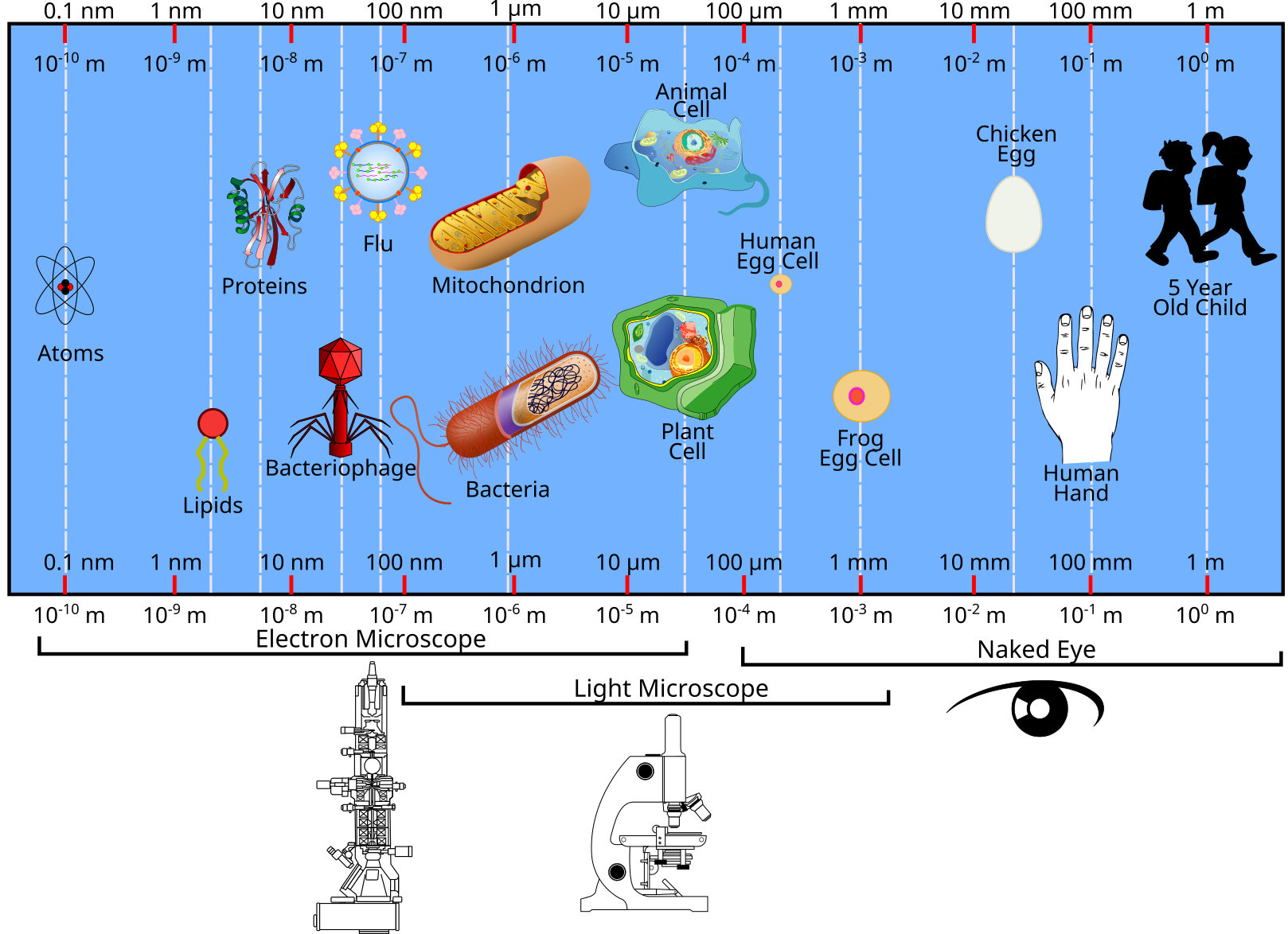
Scale

Credit: Jeremy Seto and derived from works by IgniX, Gringer, Jiver, Ninjatacoshell, Ali Zifan (CC-BY-SA)
- Discover more about scale and microscopy at this link http://learn.genetics.utah.edu/content/cells/scale/


